Microphysiological System (MPS) Related Products
Cardiac Microphysiological System MyoFlex™ Plate

Products

Trial Sales

Trial Sale Cardiac Microphysiological System MyoFlex™ Plate

Preclinical evaluation of drug-induced cardiac contractility impairment has long relied on the Langendorff perfused animal heart model. This approach, however, is limited by its inability to detect chronic toxicity and its poor human predictability.
To overcome these limitations, in vitro systems using human iPS cell-derived cardiomyocytes are being developed. In particular, 3D Engineered Heart Tissue (EHT) models have emerged as a promising next-generation platform for contractility assessment.
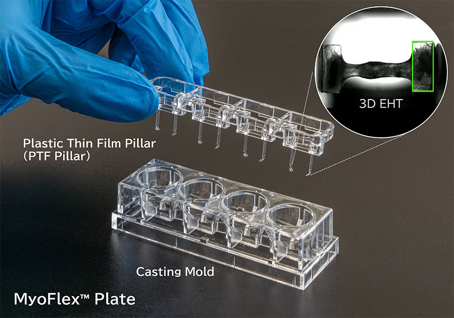
The MyoFlex™ Plate is a device dedicated for constructing EHTs from human iPS cardiomyocytes, facilitating highly accurate evaluation of drug safety and pharmacological effects.

Features

  • Plastic Thin Film Pillar (PTF Pillar): Specifically designed to minimize drug adsorption to the pillars, ensuring highly accurate toxicity assessment.
  • Uniquely designed film pillar shape: This system enables highly sensitive evaluation of contractile forces in 3D EHTs.
  • Reduce the time required to create 3D EHTs.: Our specialized molded containers are coated with an ultra-low cell attachment surface, so that gel-containing 3D EHTs can be released smoothly and easily.
MyoFlex™ Plate’s promotional video
Workflow for 3D engineered heart tissue construction using MyoFlex™ Plate
  • Preparation: Prepare a cell suspension using a fibrinogen-containing medium.
  • Mixture: Mix a small amount of thrombin solution into the fibrinogen-containing cell suspension, and add the mixture to the MyoFlex™ Plate casting mold.
  • Formation: Incubate the plate in an incubator for approximately 2 hours to form a gel-like structure.
  • Maturation: Transfer the PTF pillars to a 24-well plate containing cell culture medium, and culture them for approximately 20 days to promote maturation.
  • Observation: After the maturation period, observe that the matured 3D engineered heart tissues begin to beat spontaneously.
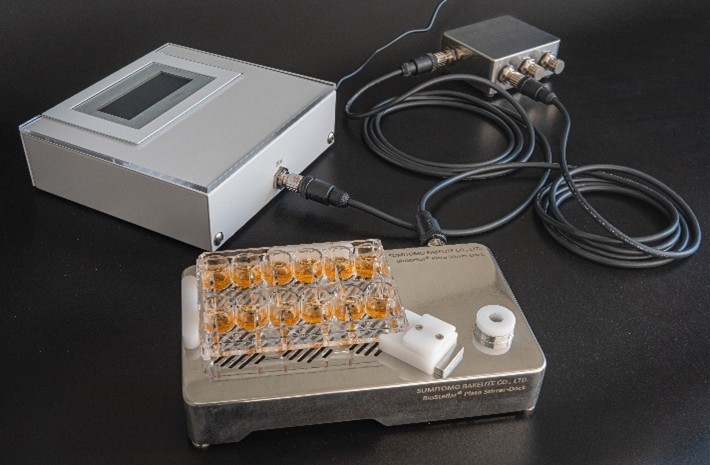
  • Quantification: Record the beating and use image analysis software to quantify the displacement of the tip of the PTF Pillar caused by contraction. Then convert this displacement into contractile force.

Application

  • Predicting drug-induced cardiotoxicity with human 3D engineered heart tissues
  • Screening of drug candidates using human 3D cardiac disease models

Application Data

Evaluation of drug response in 3D engineered heart tissue

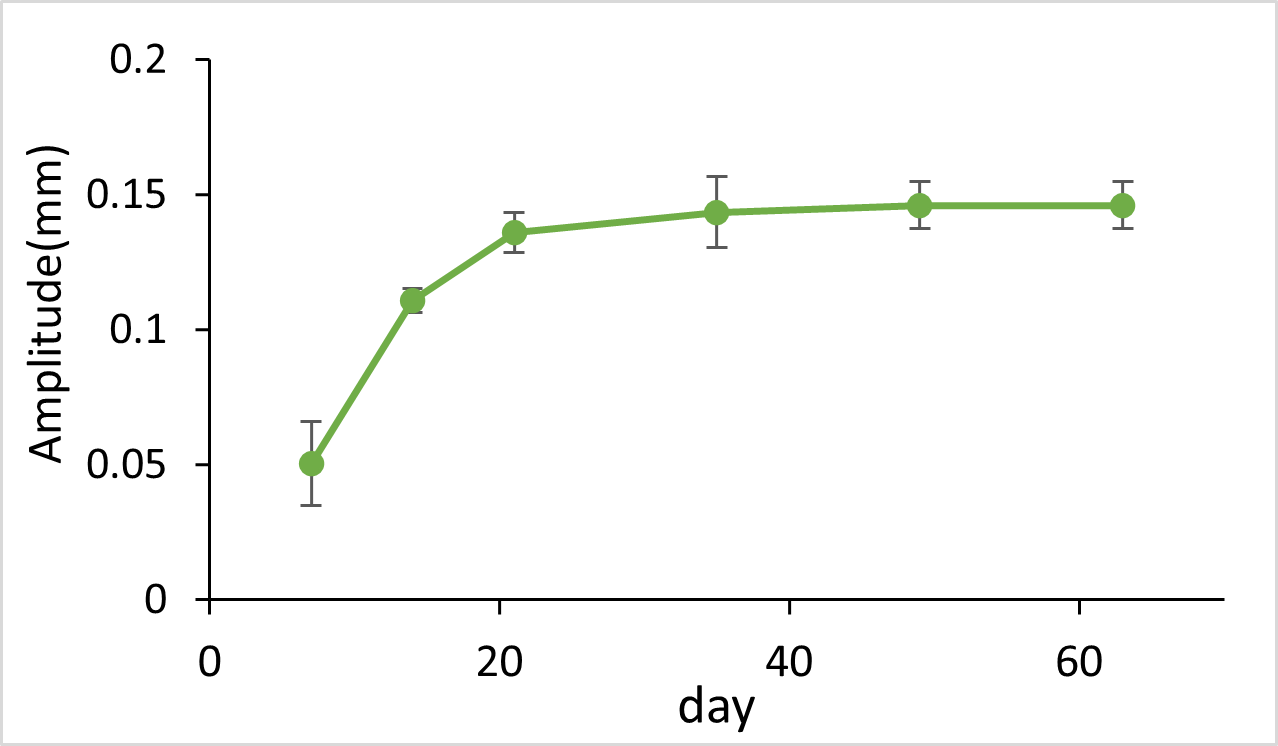
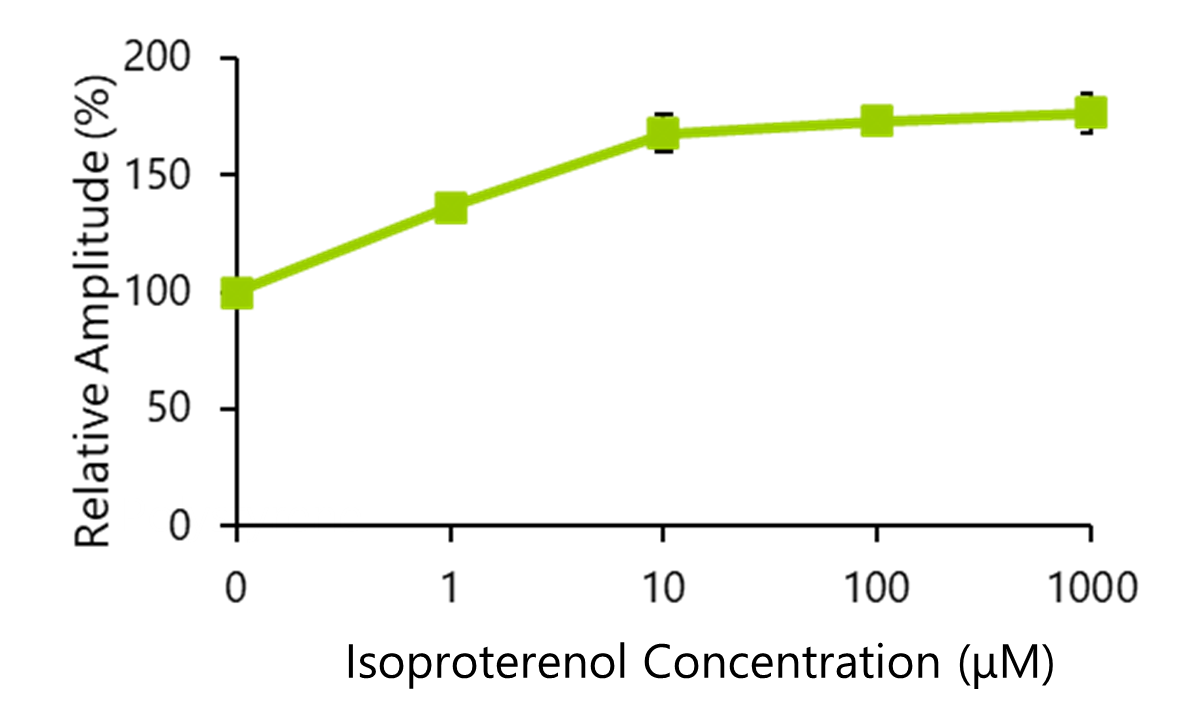
Constructed 3D heart tissues using commercially available iPS cell-derived cardiomyocytes on MyoFlex™ Plate. Evaluated the changes in contractile force associated with maturation, as well as the response (contractility and Beats per Minute: BPM) to the β-adrenoceptor agonist, Isoproterenol.

Fig.1 Contractile force change associated with the maturation of 3D heart tissue

Fig.1 Contractile force change associated with the maturation of 3D heart tissue

The contractile force increased and stabilized over the first 21 days, and the 3D heart tissue was successfully maintained for up to 63 days while sustaining its contractile strength.

Response to Isoproterenol*

Isoproterenol: The β-adrenergic receptor agonist is known as a medication that increases heart rate and myocardial contractility.

Fig.2 Changes in relative contractile force in response to Isoproterenol

Fig.2 Changes in relative contractile force in response to Isoproterenol

Fig.3 Changes in BPM in response to Isoproterenol

Fig.3 Changes in BPM in response to Isoproterenol

Both the contractile force and BPM of the 3D heart tissue exhibited a dose-dependent increase in response to Isoproterenol concentrations.

Pre-dose

Post-dose

Specifications

Product Number Product Name Contents Remarks Price
BS-X9609 MyoFlex™ Plate 3 units/case Unit Configuration
 - PTF Pillar 3 u
 - Casting mold 3 u
 - Lid 3 u
Ask

For any inquiries, quotes, or orders for BS-X9609, please contact us via the inquiry form

Publications

  • A polystyrene-film-based device for engineered cardiac tissues enables accurate analysis of drug responses on contractile properties Lab on a Chip, 2025, 25, 3682-3693.
    Authors: Yuya Fujiwara et al.
    DOI: 10.1039/D4LC00691G

Call or email us about our Division/Department.

TEL:
+81-3-5462-4831
FAX:
+81-3-5462-4835

※9:00-17:40 Mon.-Fri. (JST)

Please take a look at the FAQ first.