
Chemical Direct Conversion of Human Dermal Fibroblasts to Mesenchymal Stem Cell-like Cells
and Evaluation of Their Anti-inflammatory Activity In Vivo
Kenta Yamamoto, Toshiro Yamamoto, Yoshihiro Sowa, Makoto Seki, and Osami Mazda
Stem Cell Research & Therapy 16, 597 (2025). | https://doi.org/10.1186/s13287-025-04605-x
Copyright © Authors 2024
This article is licensed under a Creative Commons Attribution 4.0 International License (CC BY).
Background
Mesenchymal stem cells (MSCs) possess multipotency and immunoregulatory functions, making them a promising cell source for regenerative medicine in cardiovascular diseases, autoimmune diseases, and inflammatory disorders. However, allogeneic MSCs are scarce, and autologous MSCs require invasive collection from patients; furthermore, their stemness declines with long-term in vitro expansion. Therefore, in this study, we attempted chemical reprogramming of human dermal fibroblasts (HDFs) into MSC-like cells (cdMSCs) using a combination of three chemical compounds: a TGF-β receptor inhibitor, an ATM inhibitor, and a ROCK inhibitor.
Research Achievements
- Changes in cell phenotype: cdMSCs exhibited strong expression of MSC surface markers (such as CD44, CD73, CD90, CD105, CD146, CD271) and showed a clear phenotypic difference from the original HDFs. Among various extracellular marker analyses (flow cytometry), the combination of TGF-β receptor inhibitor + ATM inhibitor + ROCK inhibitor (AKF) was most effective in inducing an MSC-like phenotype.
- Acquisition of multipotency: cdMSCs acquired multipotent differentiation potential into osteoblasts and adipocytes in vitro, confirmed by specific differentiation stained with Alizarin Red S and Oil Red O, respectively.
- Molecular and epigenetic changes: RNA-seq analysis revealed significant changes in the expression of genes related to TGF-β, MAPK, Hedgehog, and WNT signaling pathways in cdMSCs. Differences in DNA methylation status (CpG sites) between cell types were also observed, suggesting progression of epigenomic reprogramming.
- Evaluation of anti-inflammatory effects: In mouse models of LPS-induced acute lung injury and autoimmune arthritis, transplantation of cdMSCs contributed to attenuation of inflammation and promotion of tissue repair. Furthermore, exosomes derived from cdMSCs promoted polarization of macrophages from the M0 state to the anti-inflammatory M2 phenotype and suppressed induction of pro-inflammatory M1 macrophages.
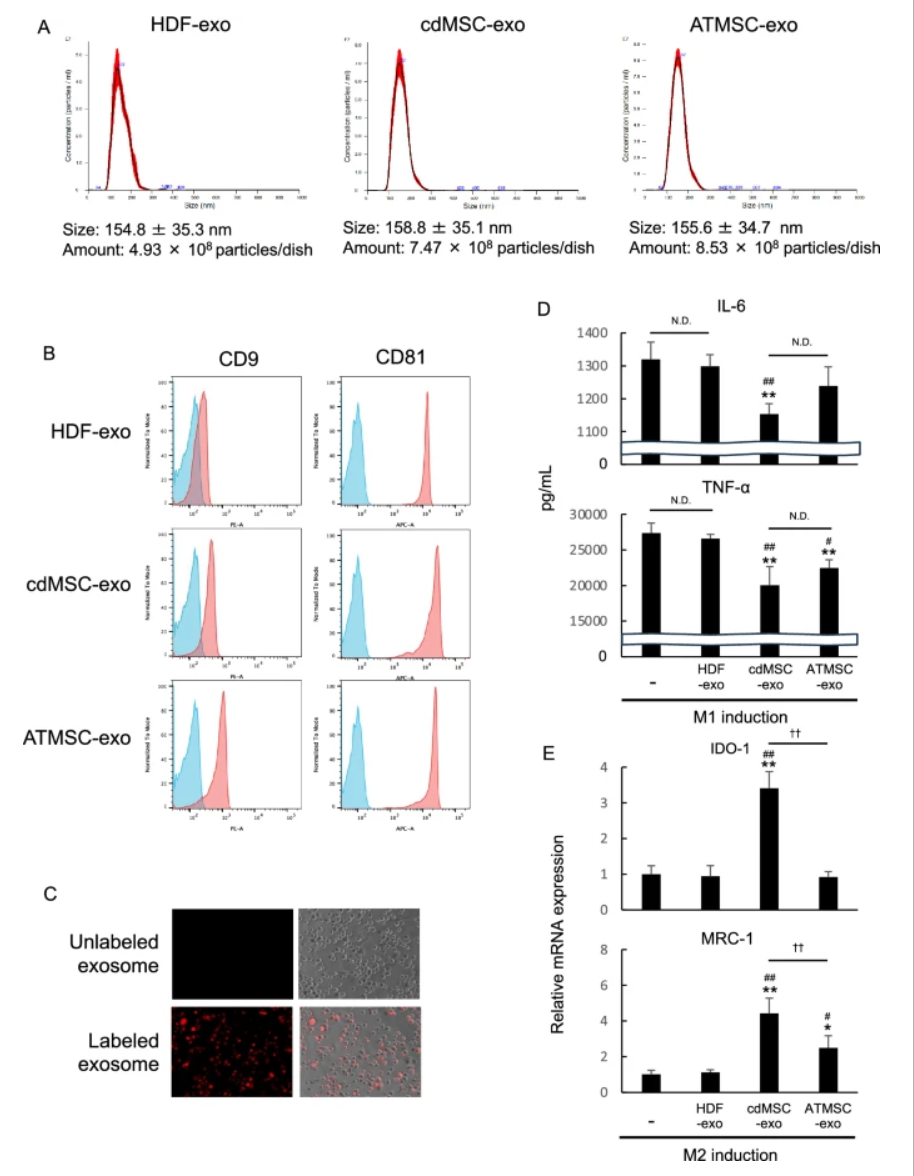
Figure 7
illustrates the characterization of exosomes derived from chemically induced MSCs (cdMSCs), including size distribution, expression of exosomal markers CD9 and CD81, and their role in promoting polarization of macrophages from pro-inflammatory M1 to anti-inflammatory M2 phenotype in vitro.
* For details, please refer to the paper
PROTEOSAVE™ Used in This Paper
In the preparation stage of exosomes in this paper, the protein low-adsorption products PROTEOSAVE™ was used. With its special coating treatment, PROTEOSAVE™ minimizes loss of protein and cell caused by their surface adsorption.
The application note regarding exosomes is available here.