Modeling embryo-endometrial interface recapitulating
human embryo implantation
Shun Shibata, Shun Endo, Luis A. E. Nagai, Eri H. Kobayashi, Akira Oike, Norio Kobayashi, Akane Kitamura, Takeshi Hori, Yuji Nashimoto, Ryuichiro Nakato,
Hirotaka Hamada, Hirokazu Kaji, Chie Kikutake, Mikita Suyama, Masatoshi Saito, Nobuo Yaegashi, Hiroaki Okae, and Takahiro Arima
Sci. Adv. 10, eadi4819 (2024). | https://doi.org/10.1126/sciadv.adi4819
Copyright © Authors 2024
This article is licensed under a Creative Commons Attribution 4.0 International License (CC BY).
Background
The early stage of human pregnancy initiation is marked by embryo implantation into the uterine endometrium; however, the underlying mechanisms remain largely elusive due to ethical restrictions and technical challenges. In particular, the 3D architecture of endometrial epithelial, stromal, and endothelial cells and their functional interactions with the embryo remain poorly understood. Traditional 2D culture models and simple coculture systems have failed to replicate in vivo tissue architecture, hormone responsiveness, and intricate cell-cell interactions.
Research Achievements
In this study, the authors developed hormone-responsive apical-out endometrial organoids (AO-EMO) that recapitulate the in vivo architecture of endometrial tissue. AO-EMO exhibited an outwardly oriented apical surface, dense stromal cells, and a self-assembled endothelial network with enhanced maturation and secretory functions upon hormonal stimulation. Coculturing AO-EMO with human embryonic stem cell-derived blastoids established a 3D feto-maternal assembloid system which recapitulated crucial implantation stages, including apposition, adhesion, and invasion. Invasion and fusion with syncytial cells and endometrial stromal cells were validated in this model using human blastocysts. Using human blastocysts, they demonstrated their adhesion onto AO-EMO surfaces and invasion into epithelial cells, enabling detailed investigation of cellular fusion and interactions during implantation.
This model, faithfully recapitulating human embryo implantation processes, This system provides a novel experimental platform to dissect the complex biochemical and physical interactions at the embryo-maternal interface, supplying insights for advancing reproductive medicine.
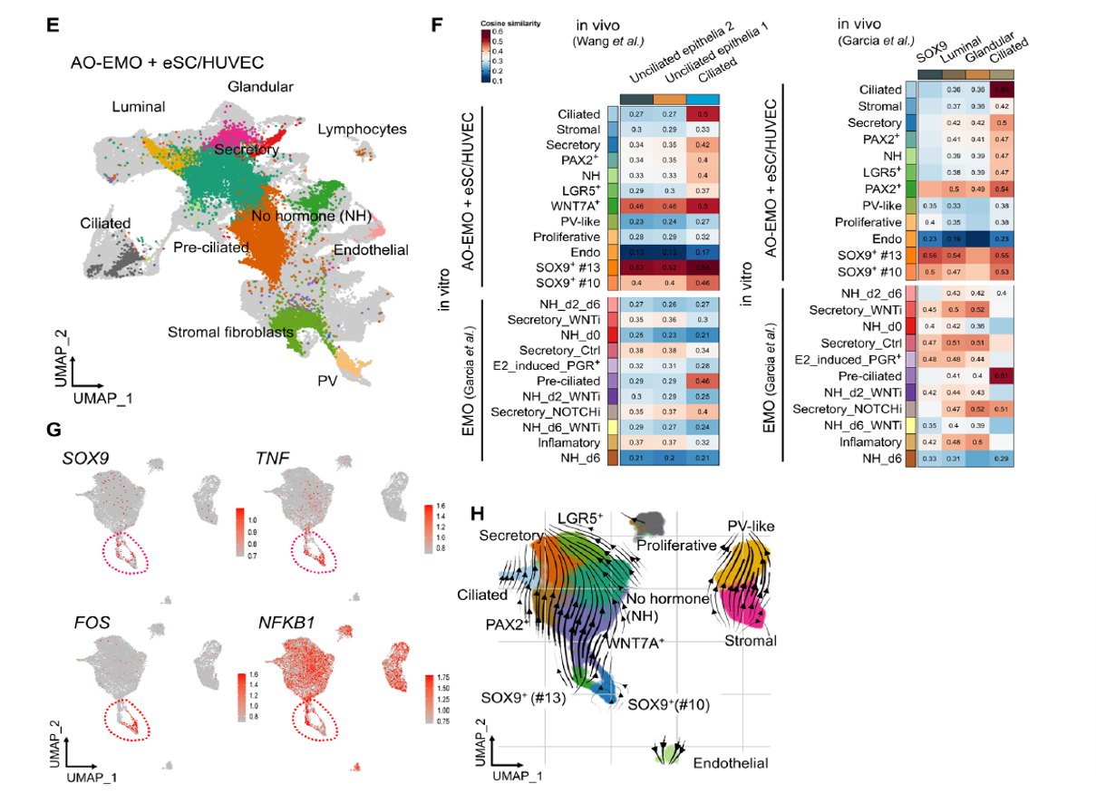
Figure 3 E-H (Multicellular integration into the 3D model and scRNA-seq analysis):
E and F: Fluorescence microscopy images showing fusion of syncytiotrophoblast cells derived from blastoid embryos on AO-EMO, forming multinucleated cells.
G and H: Heatmaps and clustering analyses of gene expression profiles, illustrating the distribution of high-quality cell populations used for analysis.
Use of STEMFULL™ in This Research
STEMFULL™ was utilized during single-cell RNA sequencing (scRNA-seq) sample preparation to handle cell suspensions. It minimized cell adhesion and loss, thereby contributing to the acquisition of high-quality scRNA-seq data.
* For details, please refer to the paper
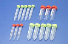
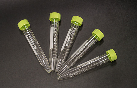
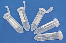
| Cat # | Product name | Material | Capacity | Packaging |
|---|---|---|---|---|
| MS-90150 | STEMFULL™ | Main body: PET Lid: Polyethylene |
15 mL | 5 pieces per pack 100 pieces per case |
Remark
- Temperature rage for use: -80°C-40°C
- Upper strength of centrifuge: 4,640G
(In-house data: Rotation time 10 min, Use of Swing rotor, rubber) - Radiation sterilized
- Storage: Room temperature. Expiration: 2 years after production