High-throughput differentiation of human blood vessel organoids reveals overlapping and distinct functions of the cerebral cavernous malformation proteins
Dariush Skowronek, Robin A. Pilz, Valeriia V. Saenko, Lara Mellinger, Debora Singer, Silvia Ribback, Anja Weise, Kevin Claaßen, Christian Büttner,
Emily M. Brockmann, Christian A. Hübner, Thiha Aung, Silke Haerteis, Sander Bekeschus, Arif B. Ekici, Ute Felbor & Matthias Rath
Angiogenesis 28, 32 (2025). | https://doi.org/10.1007/s10456-025-09985-5
Copyright © Authors 2024
This article is licensed under a Creative Commons Attribution 4.0 International License (CC BY).
Background
Cerebral cavernous malformations (CCMs) are leaky vascular lesions in the brain and spinal cord that can cause seizures or intracranial hemorrhages ICH. CCM is one of the most common cerebrovascular diseases (a prevalence of approximately 1 in 200). Although most CCMs are sporadic, there is also a familial form of CCM with three identified disease genes: CCM1, CCM2, and CCM3. However, it remains unclear why CCM3 mutations often lead to a more aggressive phenotype than CCM1 or CCM2 variants. Currently, no pharmacological therapy has yet been approved for clinical use, leaving surgery as the only option for patients with severe symptoms. To accelerate drug discovery, there is a growing need for experimental systems that faithfully recapitulate human disease and technologies compatible with high-throughput screening approaches.
Research Achievements
In this study, the authors established a protocol for high-throughput differentiation of blood vessel organoids from hiPSCs in a 96-well format, uncovering both shared and distinct functions of the CCM proteins through CCM1, CCM2, and CCM3 knockouts combined with single-cell RNA sequencing, and high-content imaging. While there was a significant overlap of differentially expressed genes in fibroblasts across all three knockout conditions, each knockout also led to specific gene expression patterns in neuronal, mesenchymal, and endothelial cell populations, respectively. Notably, mosaic blood vessel organoid analysis visualized the abnormal expansion of CCM1 and CCM3 knockout cells, whereas CCM2 knockout cells showed even reduced proliferation. This observation may help to explain the less severe clinical course in individuals with a pathogenic variant in CCM2. Finally, the excellent scalability of blood vessel organoid differentiation in a 96-well format further supports their use in high-throughput drug discovery.
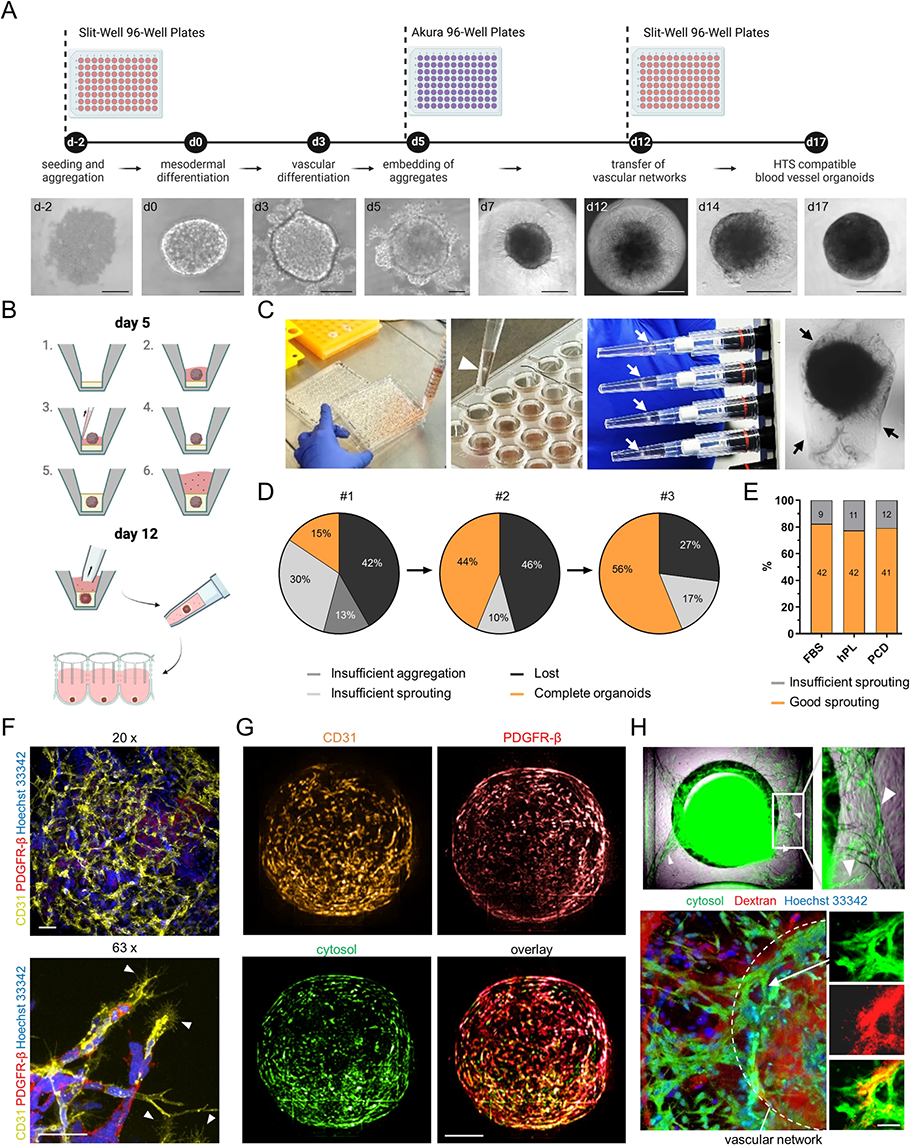
High-throughput (HT)-compatible and nearly xeno-free synthesis of vascular networks and blood vessel organoids from fluorescently tagged human induced pluripotent stem cells (hiPSCs).
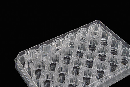
PrimeSuface™ Used in This Paper
In this study, the PrimeSurface™ Slit-well Plate 96S was used in two key steps: using the Slit-well plates for hiPSC aggregate formation allowed efficient medium; and on day 12, the matrix plugs with the vascular networks were transferred from the gel to individual wells of the PrimeSurface™ Slit-well Plate 96S by pipetting and use it to dissolve excess matrix instead of manual extraction.