
Modeling Brain Metastasis Using BioStellar™ Plate

Introduction
The blood-brain barrier (BBB) is a highly dynamic interface that tightly regulates molecular exchange between the bloodstream and the central nervous system. Conventional in vitro models often lack physiological relevance because they fail to reproduce the BBB’s complex multicellular architecture, biomechanical forces, and dynamic microenvironment. BioStellar™ Plate overcomes these limitations by enabling controlled flow, physiologically relevant shear stress, and co-culture with supporting cells such as normal human astrocytes.
In this application note, a comprehensive overview of BioStellar Plate is presented, highlighting its robustness and integration into BBB on-chip workflows, including validation of astrocyte-driven enhancement of MDA-MB-231 BR cell invasion mediated by astrocytes and their conditioned media.
Materials and Methods
• Cell Lines and Culture Media
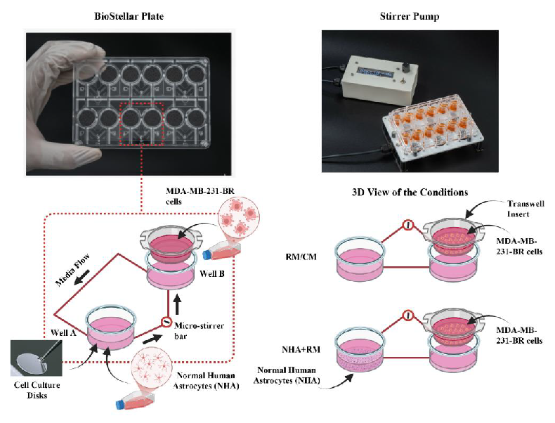
To model the multistep process of brain metastasis, MDA-MB-231 BR cells were seeded into the upper inserts (vascular channel) at 1,000 cells per insert, while NHAs were seeded at 30,000 cells per well.
This configuration enabled investigation of key metastatic processes, including adhesion, transmigration, and invasion into the brain parenchyma, within a three-dimensional, perfusable system that closely mimics the in vivo BBB.
Three media conditions were evaluated: regular media (RM), conditioned media (CM), and RM supplemented with NHAs (RM + NHA). Conditioned media was collected from confluent NHA cells cultured in regular media for 48 hours.
• Migration Assayusing Transwell and BioStellar™ Plate
Experiments were first performed using a standard transwell migration assay, in which media was changed manually every day for both inserts and bottom wells. For the RM and CM conditions, MDA-MB-231 BR cells were seeded into the insert while the respective media was added simultaneously to the bottom well. For the RM + NHA condition, both MDA-MB-231 BR and NHA cells were seeded concurrently and incubated for 24 hours prior to the first media change.
The same seeding and media change procedures were followed for BioStellar™ Plate. For the RM and CM conditions, MDA-MB-231 BR cells were allowed to adhere for 2 hours before the magnetic stirrer motor base was activated. For the RM + NHA condition, cells were similarly allowed to adhere for 2 hours prior to rotor activation. Conditioned media was applied to both wells at each media change. The magnetic stirrer motor base was operated at 5,000 rpm throughout the experiment.
• Sample Collection and Staining
At 24, 48, and 72 hours, inserts were removed and cells remaining in the upper chamber were swabbed out using a cotton tip applicator. Each insert was fixed with 70% ethanol for 15 minutes, air-dried for 4 hours, and subsequently stained with 1% toluidine blue for 10 minutes. Inserts were then washed three times with 1× dPBS and allowed to dry completely before imaging.
• Imaging and Quantification
Inserts were imaged using an EVOS M5000 microscope (ThermoFisher AMF5000). Three regions per insert were imaged, and cells were counted manually. Cell counts were averaged across the three regions for each replicate and subsequently plotted using GraphPad Prism 10. Statistical analysis was performed using a two-way ANOVA with Tukey’s multiple comparisons test.
Results
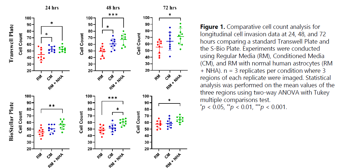
Cell counts generally increased from 24 to 72 hours across all media conditions in both the transwell plate and BioStellar™ Plate, indicating that both environments support ongoing cell invasion. The RM + NHA condition consistently yielded the highest cell counts compared to RM and CM, with the most statistically significant difference observed at 48 hours in both plate types (p < 0.001), suggesting that the addition of NHAs provides a robust invasive advantage.
While a significant difference between the RM and RM + NHA conditions was observed in both platforms, only BioStellar™ Plate demonstrated a significant difference between the RM + NHA and CM conditions at 48 hours. By 72 hours, the statistical significance between groups began to diminish across both platforms.
Conclusions
The use of conditioned media demonstrated a consistent enhancement of cell invasion, showing a reproducible increase in cell counts over the baseline RM across both the transwell and BioStellar™ Plate platforms. Significantly higher cell yields were achieved at the 24-hour and 48-hour marks under the CM condition, demonstrating that while RM + NHA supplementation provides the maximum invasive advantage, CM promotes invasion compared to standard media alone.
BioStellar™ Plate shows great potential for in vitro cell metastasis applications and is expected to be widely adopted by researchers in the fields of cell biology, tissue engineering, and biomedical applications requiring a physiologically balanced growth environment.
“Our collaborative work with SB using BioStellar™ Plate has revealed exciting potential in this next-generation microphysiological system. The platform’s ability to seamlessly support co-culture configurations, combined with its ease of operation, truly sets it apart from other MPS we have worked with to date. We look forward to exploring the many possibilities this technology has to offer.”
— Sofia D Merajver, MD, PhD, University of Michigan Medical School, Collaborative Researcher
Data contribution
Product information
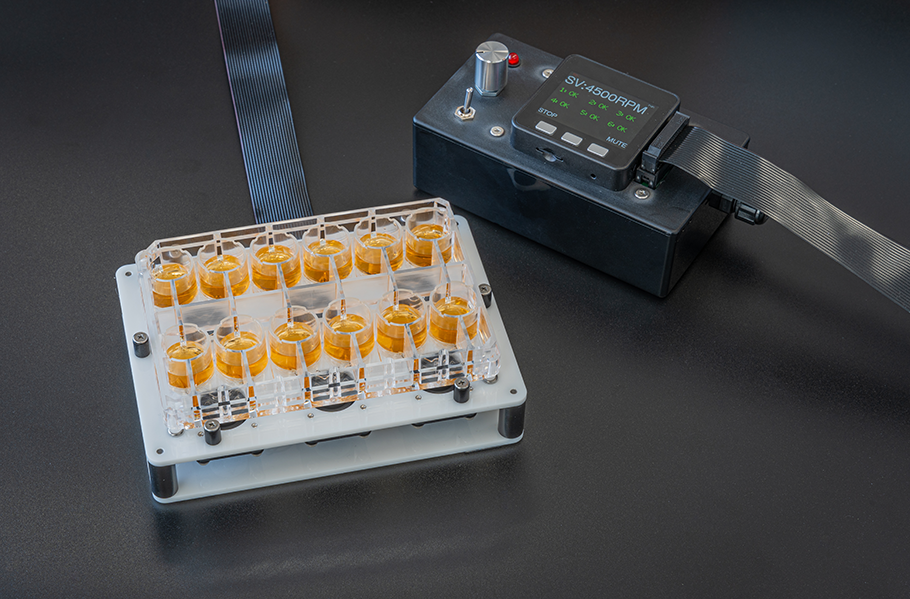
The BioStellar™ Plate is a multi-organ MPS designed for easy perfusion culture by rotating an integrated micro-stirrer bar with a dedicated device. By combining it with commercially available cell culture inserts, up to four types of cells can be co-cultured simultaneously. It features an open system structure with well sizes equivalent to those of a 24-well plate, ensuring operability comparable to standard culture equipment. Additionally, the plate does not use PDMS, resulting in minimal drug adsorption and reduced loss during evaluation.
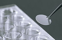
The CellDesk LF is a plastic coverslip designed to achieve low background fluorescence. It is surface-treated for adherent cells, making it suitable for cell fixation and specimen preparation. Its low autofluorescence properties make it ideal for immunochemical staining and observing intracellular localization using fluorescent markers.
| Product Number | Product Name | Contents | Price |
|---|---|---|---|
| BS-X9607 | BioStellar™ Plate | 2 plates / case | ASK |
| MS-92132 | Cell Desk LF1 | 240 (24×10) / CASE | ASK |
* For any inquiries, quotes, or orders regarding the products, please feel free to contact us directly.
E-mail: s-bio_inquiry@ml.sumibe.co.jp