
Application: Interaction between Immune cells and a 3D Skin Model Using BioStellar™ Plate

Introduction
Understanding skin immune responses is crucial for studying allergic dermatitis, infectious diseases, and developing new treatments. Traditional 2D cell culture systems using single cell types cannot fully replicate the complex intercellular reactions of the body. Microphysiological Systems (MPS) are increasingly used as advanced co-culture platforms designed to better mimic in vivo environment. Sumitomo Bakelite Co., Ltd.’s BioStellar™ Plate, a multi-organ MPS with perfusion capabilities, offers a more realistic reproduction of these interactions. In this study, we present a case study investigating the interaction between skin and immune cells using the BioStellar™ Plate. THP-1 cells were co-cultured with LabCyte EPI-MODEL24 (EPI-MODEL), a 3D human skin model developed by Japan Tissue Engineering (J-TEC), under perfusion conditions to mimic in vivo dynamics. After the culture, specific surface biomarkers expressed on THP-1 cells were analyzed by flow cytometry and the secreted inflammatory cytokine, interleukin 8 (IL-8) in the supernatant was measured using ELISA. The results highlight the potential of our MPS as a physiologically relevant model for investigating skin inflammation and immune responses.
Materials and Methods
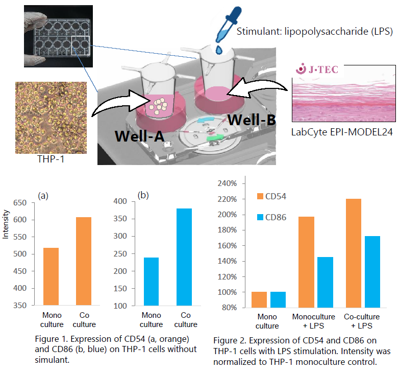
- Inserts seeded with THP-1 cells (1x10⁵ cells/insert) were transferred to well A of a BioStellar™ Plate, while well B was loaded with inserts containing EPI-MODEL. The cells were co-cultured under erfusion flow at 4,500 RPM for 48 hours.
To assess the effect of immune stimulation, lipopolysaccharide (LPS) was additionally added to the culture medium at a final concentration of 50 ng/mL under the same co-culture conditions. Under both unstimulated and LPS-stimulated conditions, THP-1 monocultures were included as controls. After 48 hours, THP-1 cells were harvested and surface biomarkers, CD54 and CD86 were analyzed by flow ytometry. - To evaluate the inflammatory response and the communication between THP-1 cells and skin cells, the supernatant was collected from the basal compartment of the inserts in well B following culture, and L-8 levels were measured by ELISA.
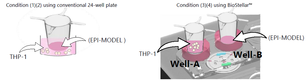
Furthermore, to evaluate the effect of perfusion, IL-8 concentrations were compared across four conditions: (1) THP-1 monoculture in a conventional 24-well plate, where THP-1 cells were seeded directly on the well bottom, (2) THP-1 and EPI-MODEL co-culture in a conventional 24-well plate, (3) Insert seeded THP-1 on the BioStellar late, and (4) Insert seeded THP-1 and EPI-MODEL co-culture on the BioStellar™ Plate.
Results 01
Surface biomarkers CD54 and CD86 expressed on THP-1 cells were analyzed after THP-1 cells and EPI-MODEL were co-cultured on the BioStellar™ Plate for 48 hours with or without LPS.
Without stimulation, surface expression of both CD54 and CD86 on THP-1 cells was slightly elevated in co-culture with EPI-MODEL compared with monoculture (Figure1). LPS stimulation led to further upregulation of both biomarkers, with the most pronounced increases observed in the THP-1 + Skin co-culture condition, reaching ~220% for CD54 and ~170% for CD86 relative to monoculture controls (Figure 2). These results suggest that interaction between THP-1 cells and skin cells was established in our MPS platform, enhancing the LPS-induced immune activation of THP-1 cells.
Results 02
To evaluate inflammatory responses, THP-1-Skin communication, and the effect of perfusion, IL-8 levels in the culture supernatant were measured by ELISA across four conditions below:
| Conditionr | Device | Cell | IL-8 (pg/mL) | medium volume |
|---|---|---|---|---|
| (1) | 24-well plate | THP-1 | <1.37 pg/mL | 500μl |
| (2) | 24-well plate | EPI-MODEL (insert) THP-1 (plate) |
21.4 (pg/mL) | 500μl |
| (3) | BioStellar™ Plate | THP-1 (Well-A) | <1.37 pg/mL | 1200μl (insert 150μl) |
| (4) | BioStellar™ Plate | EPI-MODEL (Well-B) THP-1 (Well-A) |
47 (pg/mL) | 1200μl (insert 150μl) |
IL-8 levels in THP-1 monoculture were below the limit of detection under both the conventional 24-well plate and the BioStellar™ Plate conditions. In contrast, IL-8 secretion was detectable when EPI-MODEL was present in coculture; condition 2 (conventional 24-well plate) produced 21.4 pg/mL, whereas condition 4 (BioStellar™ Plate) yielded a markedly higher level of 47 pg/mL. These results indicate that the presence of skin cells promotes IL-8 secretion, and that perfusion culture on the BioStellar plate further enhances this inflammatory response compared to conventional static culture.
Conclusion
This study demonstrated that co-culture of THP-1 cells with LabCyte EPI-MODEL24 under perfusion conditions on the BioStellar™ Plate enhanced both immune cell activation, evidenced by increased CD54 and CD86 expression, as well as inflammatory cytokine secretion, reflected by elevated IL-8 levels compared with conventional static culture. These findings highlight the BioStellar™ plate a powerful platform to study complex interactions between immune cells and skin tissue under controlled conditions. By leveraging perfusion, this system enables the observation of dynamic processes, such as metabolite exchange and immune–slkin crosstalk, which are critical for understanding physiological and pathological responses. Furthermore, the seamless compatibility of the BioStellar™ Plate with 3D skin models such as LabCyte EPIMODEL24 allows for more realistic and direct modeling of skin-related immune responses, making it a valuable tool for inflammation research, drug testing, and the therapeutic development.
Product information
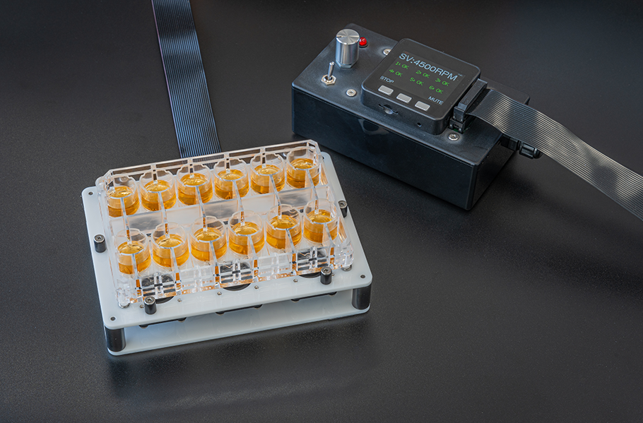
The BioStellar™ Plate is a multi-organ MPS designed for easy perfusion culture by rotating an integrated micro-stirrer bar with a dedicated device. By combining it with commercially available cell culture inserts, up to four types of cells can be co-cultured simultaneously. It features an open system structure with well sizes equivalent to those of a 24-well plate, ensuring operability comparable to standard culture equipment. Additionally, the plate does not use PDMS, resulting in minimal drug adsorption and reduced loss during evaluation.
The CellDesk LF is a plastic coverslip designed to achieve low background fluorescence. It is surface-treated for adherent cells, making it suitable for cell fixation and specimen preparation. Its low autofluorescence properties make it ideal for immunochemical staining and observing intracellular localization using fluorescent markers.

| Product Number | Product Name | Contents | Price |
|---|---|---|---|
| BS-X9607 | BioStellar™ Plate | 2 plates / case | ASK |
| MS-92132 | Cell Desk LF1 | 240 (24×10) / CASE | ASK |
* For any inquiries, quotes, or orders regarding the products, please feel free to contact us directly.
E-mail: s-bio_inquiry@ml.sumibe.co.jp